
Multimodal microscopy
Members involved
Sylvain Rivet, Matthieu Dubreuil, Yann Le Grand, Gaël Leroux (technician).
Objectives
Implementing multimodal and polarimetric imaging (Mueller, SHG, TPEF) on a laser scanning microscope. Applications in the fields of biophotonics (diagnosis, animal and plant tissue imaging, extracellular matrix imaging…) and of material science (liquid crystals, thin films...).
Description
Custom made imaging modalities were (and are being) developed from both commercial (Olympus BX51WI-FV300) and modular laser scanning microscopes to provide various contrasts such as those based on Mueller polarimetry, second harmonic generation (SHG) and two-photon excitation fluorescence (TPEF).
Mueller microscopy
The first laser scanning microscope able to acquire a Mueller image in a single scan was developed by our group. The Mueller polarimeter uses a swept laser source at 100kHz (see section spectral domain Mueller polarimetry), which is compatible with the pixel dwell-time of the microscope. The acquisition of channeled spectra was synchronized with galvo scanners and PSG/PSA blocks were integrated into the microscope optical path. The microscope currently works in transmission a will be extended to the reflection geometry. Real-time display of images is currently under development.
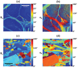
408×512 Mueller images of (a), (b) a human liver biopsy, and (c), (d) a section of gabbro rock. (a), (c) Linear retardance RL, and (b), (d) its orientation αR. Image field: 1.75×1.75 mm.
SHG/TPEF microscopy
A femtosecond oscillator (Verdi-Mira - Coherent) is used as a laser source. Custom made SHG (transmission and backscattering) and TPEF (descanned and non-descanned in backscattering) detection paths were implemented on scanning confocal microscope. Several biomedical studies were conducted in collaboration with biologist and physician groups.
- Collagen imaging and quantification for liver fibrosis diagnosis
SHG image of a F4 human liver specimen
- Collagen and elastin imaging and quantification in vessel walls for evaluation of atherosclerosis
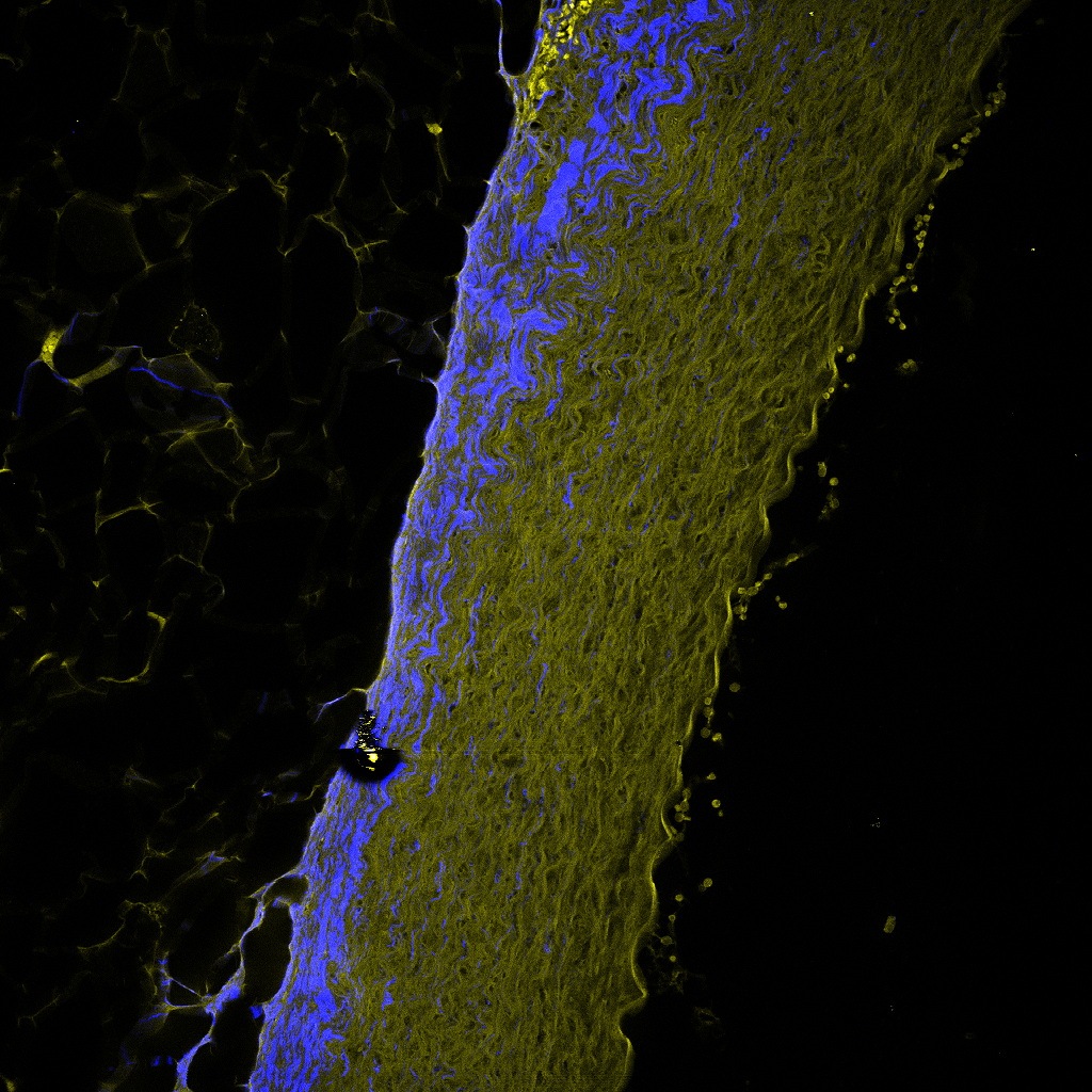
Combined SHG (blue) and TPEF (yellow) image of a rabbit vessel wall
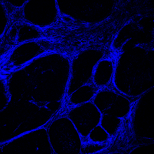
- Study of re-innervation of human skin
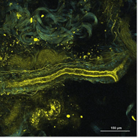
Nerve (yellow) and collagen (blue) fibers in a re-innervated human skin model
as imaged by coupled TPEF and SHG microscopies
- Polarimetric SHG imaging of rat muscle in sepsis
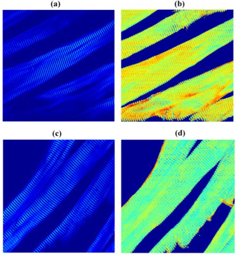
(a,c) Circularly polarized SHG image (b,d) anisotropy parameter SHG image
of rat EDL muscle in control (a,b) and septic (c,d) conditions
PhD-thesis
A. Le Gratiet, "Développement d'un polarimètre de Mueller à codage spectral utilisant une swept-source : application à la microscopie à balayage laser". University of Brest - UBO (2016). Supervisor : Y. Le Grand and S. Rivet.
D. Sevrain, "Développements en microscopie non linéaire cohérente et incohérente et applications". University of Brest - UBO (2013). Supervisor : Y. Le Grand.
Selection of publications
M. Dubreuil, F. Tissier, L. Le Roy, J.P. Pennec, S. Rivet, M.-A. Giroux-Metges, Y. Le Grand "Polarization-resolved second harmonic microscopy of skeletal muscle in sepsis" Biomed. Opt. Express 9(12), 6350-6358 (2018).
A. Le Gratiet, S. Rivet, M. Dubreuil and Y. Le Grand, "Scanning Mueller polarimetric microscopy", Opt. Letters 41(18), 4336-4339 (2016).
F. Tissier, Y. Mallem, C. Goanvec, R. Didier, T. Aubry, N. Bourgeois, J-C Desfontis, M. Dubreuil, Y. Le Grand, J. Mansourati, K. Pichavant-Rafini, E. Plee-Gautier, P. Roquefort, M. Theron, M. Gilard, "A non-hypocholesterolemic atorvastatin treatment improves vessel elasticity by acting on elastin composition in WHHL rabbits", Atherosclerosis 2514, 70-77 (2016).
D. Sevrain, M. Dubreuil, G.E Dolman, A.M. Zaitoun, W.L. Irving, I.N. Guha, C. Odin, Y. Le Grand "Evaluation of area-based collagen scoring by nonlinear microscopy in chronic hepatitis C-induced liver fibrosis", Biomed. Opt. Express 6(4):1209–1218 (2015).
D. Sevrain, Y. Le Grand, V. Buhé, C. Jeanmaire, G. Pauly, J-L. Carré, L. Misery, N. Lebonvallet, "Two-photon microscopy of dermal innervation in a human re-innervated model of skin", Experimental dermatology, Volume 22, Issue 4, pages 290–291 (2013).
D. Sevrain, M. Dubreuil, A. Leray, C. Odin, Y. Le Grand, "Measuring the scattering coefficient of turbid media from two-photon microscopy" Opt. Express 21, 25221-25235 (2013).
M. Dubreuil, P. Babilotte, L. Martin, D. Sevrain, S. Rivet, Y. Le Grand, G. Le Brun, B. Turlin, B. Le Jeune "Mueller matrix polarimetry for improved liver fibrosis diagnosis", Opt. Letters, Vol. 37, Issue 6, pp.1061-1063 (2012).
Fundings
CPER Sophie Photonique 2015-2020 (200k€) : femtosecond laser (Vision II - Coherent) + scanning microscope (Thorlabs) and accessories.
Collaborations
- Applied Optics Group (AOG), University of Kent, UK.
- Laboratoire ORPHY, Université de Bretagne Occidentale, France.
- Laboratoire des neurosciences de Brest (LNB), Université de Bretagne Occidentale, France.
- Institut de Physique de Rennes (IPR), CNRS/Université de Rennes 1, France
- Nottingham Digestive Diseases Biomedical Research Unit, Nottingham, UK.
